Dendritic spines are critical for synaptic plasticity associated with learning and memory. Because alterations in spine number or shape are correlated with cognitive dysfunction in various neurological disorders, they are of significant interest as a subcellular substrate for anesthetic action. Long-term cognitive and amnestic changes have been described following anesthesia, suggesting a role for spine plasticity in the enduring, deleterious effects of anesthetics.

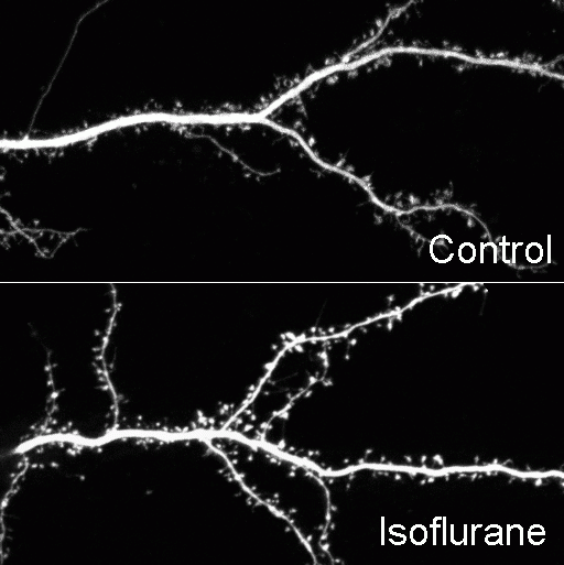
Confocal time-lapse images of GFP labeled hippocampal neurons (21 DIV) showing dendritic arbor of control (top) and isoflurane-treated (below) neurons. (From Platholi et al., 2014).

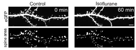
Confocal time-lapse images of GFP labeled hippocampal neurons (21 DIV) showing dendritic arbor of control and isoflurane-treated neurons (top) and corresponding thresholded overall spine area after dendrite subtraction (below). Scale bar = 5μm. (From Platholi et al., 2014).